
Ringkasan Eksekutif
Bandingkan alas meja operasi serat karbon vs standar untuk pencitraan C-arm, O-arm, dan CT intraoperatif. Mencakup radiolusensi, kepatuhan IEC 60601-2-46, dan trade-off beban.
Every orthopedic surgeon has experienced it: the C-arm swings into position, the technician fires a shot, and the image comes back with a dark band across the femoral neck. The cause? A standard phenolic-resin table top with a steel reinforcement channel sitting directly in the beam path. The team repositions the patient, fires again, and burns another 15 seconds of fluoroscopy time. Multiply that by forty cases a month and you have measurable radiation overexposure, longer OR turnover, and frustrated imaging staff.
This is the problem that radiolucent carbon fiber operating table tops solve. By replacing metal and dense composite substrates with carbon fiber reinforced polymer (CFRP), manufacturers create a patient-support surface that is largely transparent to X-rays, CT beams, and fluoroscopic radiation. The result is uninterrupted imaging access across the entire working zone, from cervical spine to distal femur, without repositioning the patient or accepting degraded image quality.
In this guide, we break down what radiolucency actually means in engineering terms, compare carbon fiber against conventional table top materials, examine compatibility with C-arm, O-arm, and intraoperative CT systems, and walk through the trade-offs between safe working load and imaging performance. Whether you are specifying tables for a new hybrid OR or upgrading an existing trauma suite, this reference will help you make an informed procurement decision. For a full overview of our product range, visit our operating tables collection.

What Does Radiolucent Mean for an Operating Table?
Radiolucent describes any material that permits ionizing radiation, specifically X-ray photons, to pass through with minimal absorption or scatter. In the context of an operating table, a radiolucent top produces no visible shadow, streak, or artifact on the resulting radiograph or fluoroscopic frame. The clinical implication is direct: the surgeon sees anatomy, not table hardware.
The physics is straightforward. X-ray attenuation follows an exponential decay governed by the material’s linear attenuation coefficient and thickness. Metals like stainless steel (density approximately 7.9 g/cm3) attenuate clinical X-ray beams almost completely at typical diagnostic energies. Carbon fiber reinforced polymer, by contrast, has a density around 1.5-1.6 g/cm3 and an atomic composition (predominantly carbon, atomic number 6) that interacts weakly with photons in the 50-120 keV range used in clinical imaging. Published radiographic analysis of fiber reinforced polymer composites confirms that carbon fiber exhibits significantly lower mass attenuation coefficients than glass or aramid alternatives at 50 keV clinical X-ray energies.
For procurement teams, the practical specification to request is “full-length radiolucent imaging zone” rather than a single radiolucent window. A table that offers only a 400 mm radiolucent section forces the surgical team to align the anatomy precisely over that window, adding setup time and limiting flexibility during complex multi-level procedures.
For where the radiolucent section sits in the five-section layout, see our five-section operating table top guide.
Procurement tip: When reviewing tender specifications, ask the manufacturer to provide attenuation test data at 70 kVp and 120 kVp. A genuinely radiolucent carbon fiber top will show less than 5% signal loss compared to an open-beam reference at both energies. Anything higher suggests hidden metal reinforcement or excessive resin content.
Carbon Fiber vs. Standard Table Top Materials
Operating table tops have historically been manufactured from three material families: stainless steel, phenolic resin (high-pressure laminate), and carbon fiber reinforced polymer. Each carries distinct implications for imaging compatibility, weight, durability, and cost. In our factory, we have produced all three configurations and can speak to the real-world differences that matter in a busy OR.
Stainless steel tops are the legacy option. They are robust, easy to fabricate, and inexpensive. However, they are completely opaque to X-rays. Any procedure requiring intraoperative imaging on a steel top demands either a separate radiolucent insert or patient transfer to a dedicated imaging table, both of which introduce workflow friction and contamination risk.
Phenolic resin tops represent a middle ground. High-pressure laminates can be formulated to be partially radiolucent, but they typically require a thickness of 12-15 mm for structural rigidity, and the resin matrix still attenuates a meaningful fraction of the beam. More critically, phenolic tops often incorporate aluminum or steel sub-frames for mounting hardware, creating localized opaque zones that appear as artifacts on lateral and oblique projections.
Carbon fiber reinforced polymer tops deliver the highest imaging performance. CFRP achieves equivalent or superior structural stiffness at roughly one-third the weight of aluminum and one-fifth the weight of steel. The material is corrosion-free, chemically stable, and withstands the aggressive enzymatic cleaners and autoclave-adjacent sterilization cycles of daily clinical use. The trade-off is raw material cost: aerospace-grade carbon fiber prepreg is significantly more expensive per kilogram than steel sheet or phenolic laminate.
| Parameter | Stainless Steel | Phenolic Resin (HPL) | Carbon Fiber (CFRP) |
|---|---|---|---|
| X-ray attenuation | Complete (opaque) | Partial (15-40% depending on thickness) | Minimal (near-transparent at clinical energies) |
| Imaging artifact risk | High (beam-hardening, streak) | Moderate (sub-frame shadows) | Negligible |
| Relative weight | Heaviest | Sedang | 60-70% lighter than aluminum equivalent |
| Corrosion resistance | Good (316L grade) | Baik | Excellent (inert polymer matrix) |
| Cleaning/sterilization tolerance | Sangat Baik | Good (edge sealing critical) | Excellent (non-porous surface) |
| Typical radiolucent zone | None (requires insert) | Partial section only | Full-length (up to 1400 mm) |
| Relative material cost | Rendah | Sedang | Tinggi |
| Best application | General surgery (no imaging) | Budget imaging suites | Hybrid OR, trauma, spine, interventional |

Imaging Compatibility: C-Arm, O-Arm, and Intraoperative CT
Not all intraoperative imaging systems place the same demands on a table top. Understanding the geometric and attenuation requirements of each modality helps you specify the correct radiolucent configuration.
C-Arm Fluoroscopy
The mobile C-arm remains the workhorse of intraoperative imaging. It projects a cone beam from below or above the patient onto a flat-panel or image-intensifier detector. For a C-arm to produce a clean image, every material between the X-ray source and the detector must be radiolucent. A carbon fiber table top allows the C-arm to shoot through the table at any angle, including steep obliques and lateral projections, without introducing metallic shadows. Standard imaging tables with carbon fiber tops provide an expanded radiolucent area, some offering up to 1900 mm (75 inches) of unobstructed imaging length, enabling full-body X-ray scans without moving the patient.
O-Arm and Intraoperative CT
O-arm systems (cone-beam CT) and dedicated intraoperative CT scanners rotate a gantry around the patient, acquiring volumetric data from 360 degrees. This places a stricter requirement on the table: the entire cross-section of the patient support must be free of high-density material, because the beam passes through the table from every azimuthal angle. Even a small steel mounting bracket that would be invisible on a single AP shot will produce streak artifacts across multiple CT slices. Full carbon fiber construction, including the side rails and locking mechanisms in the imaging zone, is essential for artifact-free 3D acquisition.
Hybrid OR Ceiling-Suspended Systems
Hybrid operating rooms combine surgical capability with fixed ceiling-mounted flat-panel detectors or robotic C-arm systems. These installations demand complete radiolucency and a table column design that does not obstruct the imaging field of view from any angle. The table pedestal geometry must be evaluated against the full range of detector travel to ensure no mechanical interference during rotational acquisitions.
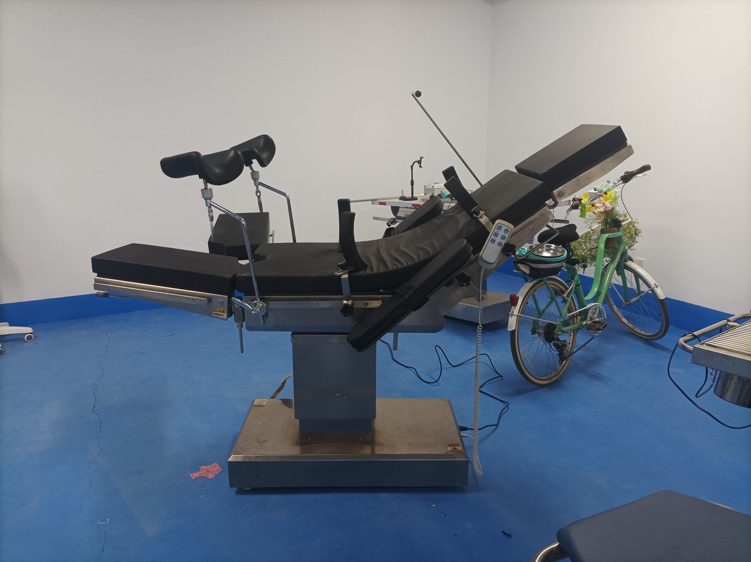
For a deeper dive into selecting tables specifically for C-arm workflows, see our Panduan meja operasi yang kompatibel dengan C-arm, which covers column geometry, lateral roll, and detector clearance in detail.
Safe Working Load vs. Radiolucency: The Engineering Trade-Off
Here is the tension that every table designer faces: the structural elements that carry patient weight are often the same elements that would ideally be radiolucent. A thicker carbon fiber laminate increases stiffness and load capacity but adds material in the beam path. Metal reinforcement brackets solve point-load problems but create imaging dead zones.
IEC 60601-2-46:2016, the international safety standard for operating tables, establishes the baseline mechanical requirements. The standard references a standard patient mass of 135 kg, with load distribution defined in IEC 60601-1 clause 9.8.3 (approximately 40.7% of load applied to the torso section). Manufacturers must demonstrate structural integrity under this load with a defined safety factor. For bariatric applications, the engineering envelope expands significantly: modern bariatric operating tables are designed to handle dynamic loads of at least 350 kg, requiring reinforced hydraulics and a solid base column to manage the enormous leverage exerted during Trendelenburg positioning.
The practical consequence for imaging tables is a weight-capacity ceiling. Most carbon fiber imaging tables support patients up to 227 kg (500 lbs), but positioning options become limited at maximum weight. The enhanced lateral roll and extended longitudinal travel that make these tables ideal for imaging also introduce structural cantilevers that reduce the maximum safe load compared to a fixed-column general surgery table.
Engineering reality: A table rated for 450 kg static load in a neutral supine position may only be rated for 250 kg in full Trendelenburg with lateral tilt. Always verify the dynamic load rating at the extreme positions your surgical team actually uses, not just the brochure headline number.
Bagi pembeli yang mengevaluasi meja operasi listrik versus hidrolik, the drive mechanism also interacts with load capacity. Electro-hydraulic systems typically deliver smoother motion under heavy loads and maintain position more reliably during extended imaging sequences, whereas all-electric systems offer finer incremental control but may have lower maximum load ratings.

Full Carbon Fiber vs. Radiolucent Section Designs
Not every surgical discipline requires full-length radiolucency. The decision between a full carbon fiber top and a hybrid design (radiolucent imaging section plus conventional sections) depends on case mix, budget, and workflow priorities.
A full carbon fiber top provides uninterrupted imaging access from head to toe. For orthopedic trauma tables, this is a non-negotiable specification: femoral nailing, tibial fixation, and pelvic fracture repair all demand fluoroscopic access across multiple anatomical zones within a single procedure. Spine surgery similarly benefits from full-length radiolucency, as pedicle screw placement at thoracic and lumbar levels requires AP and lateral shots at varying longitudinal positions.
A radiolucent-section design places a carbon fiber panel (typically 600-900 mm long) in the primary imaging zone, with phenolic or composite sections at the head and leg ends. This approach reduces material cost while still serving the majority of imaging needs. However, it introduces a constraint: if the surgical target migrates outside the radiolucent window during the procedure, the team must either reposition the patient or accept a degraded image.
- Full carbon fiber top: Best for trauma, spine, interventional radiology, hybrid OR, and any workflow where imaging field of view is unpredictable.
- Radiolucent section (partial): Suitable for elective orthopedics with predictable imaging zones, urology with occasional fluoroscopy, and budget-constrained facilities.
- Hybrid with interchangeable panels: Offers flexibility for multi-specialty ORs; carbon fiber panel swaps in for imaging cases, standard panel for general surgery.
If your facility is exploring custom configurations, our Kustomisasi meja operasi OEM guide explains how we adapt table top geometry, rail systems, and radiolucent zone length to match specific clinical protocols and imaging equipment clearances.
Compliance and Standards: IEC 60601-2-46 and Beyond
Any operating table sold into regulated markets must comply with IEC 60601-2-46:2016 (third edition), which specifies particular requirements for the basic safety and essential performance of operating tables. This standard was prepared by IEC subcommittee 62D and aligns structurally with the IEC 60601-1:2005 general standard and its Amendment 1 (2012).
For imaging-compatible tables, two additional normative references within IEC 60601-2-46 are particularly relevant:
- IEC 60601-1-3:2008 (Radiation protection in diagnostic X-ray equipment): Governs the radiation safety aspects when the table is used in conjunction with X-ray imaging systems. This collateral standard ensures that the table’s presence in the beam path does not compromise patient or operator radiation protection.
- IEC 60601-2-43 (X-ray equipment for interventional procedures): Applies when the table is integrated with fixed interventional X-ray systems, as in a hybrid OR or cath lab configuration.
Beyond electrical safety, the standard addresses mechanical hazards including stability under load, locking mechanism reliability, and protection against unintended movement. Table height adjustment ranges typically span from 650 mm to 1,050 mm from floor to table top, accommodating both seated and standing surgical teams. The standard also references IEC 60601-1-2:2014 for electromagnetic compatibility, ensuring that the table’s drive electronics do not interfere with sensitive imaging detectors.
For CE-marked devices entering the European market, compliance with IEC 60601-2-46 is assessed under the EU Medical Device Regulation (MDR 2017/745), with the manufacturer’s quality management system certified to ISO 13485. Buyers should request the manufacturer’s EC certificate and test reports as part of the tender evaluation, not merely a self-declaration of conformity.
Compliance checkpoint: Verify that the manufacturer’s IEC 60601-2-46 test report explicitly covers the radiolucent top configuration you are purchasing. Some manufacturers certify a base model with a standard top and do not re-test when swapping to a carbon fiber panel. The attenuation characteristics and mechanical load paths differ, and the certification should reflect the actual shipped configuration.
Kesimpulan
Selecting a radiolucent operating table is not simply a materials decision. It is a systems-level choice that affects imaging quality, radiation dose, OR workflow, patient safety, and long-term total cost of ownership. Carbon fiber reinforced polymer has become the industry standard for imaging-compatible table tops because it uniquely combines near-total radiolucency with structural stiffness, low weight, and clinical durability. However, the engineering trade-offs between load capacity, radiolucent zone length, and cost mean that the optimal specification varies by clinical application.
Key Takeaways:
- Radiolucency is a spectrum, not a binary. Full carbon fiber tops deliver the lowest attenuation and fewest artifacts; partial radiolucent sections are a cost-effective compromise for predictable imaging zones.
- Match the table to your imaging modality. C-arm fluoroscopy tolerates minor attenuation; intraoperative CT and O-arm systems demand full 360-degree radiolucency with zero metallic content in the imaging zone.
- Verify dynamic load ratings at extreme positions. The brochure static load number is necessary but insufficient; confirm capacity in Trendelenburg, lateral tilt, and extended longitudinal travel.
- Insist on configuration-specific certification. IEC 60601-2-46 test reports must cover the exact table top and accessory combination you are procuring, not a generic base model.
If you are specifying tables for a new build or upgrading an imaging suite, our engineering team can provide attenuation test data, load-rating matrices, and 3D clearance drawings for your specific C-arm or CT system. Explore our operating table range or request a technical consultation to match the right radiolucent configuration to your clinical workflow.
Pertanyaan yang Sering Diajukan
Apa perbedaan antara radiolusen dan radiopak pada meja operasi?
Bahan radiolusen memungkinkan sinar-X melewatinya, sehingga tidak menghasilkan bayangan pada gambar. Bahan radiopak memblokir sinar-X, tampak sebagai artefak putih atau gelap. Serat karbon bersifat radiolusen; baja tahan karat bersifat radiopak.
Dapatkah meja karbon fiber digunakan dengan semua merek C-arm?
Ya. Radio-lusensi serat karbon bersifat berbasis material, bukan spesifik merek. Setiap C-arm bergerak atau yang digantung di langit-langit mendapat manfaat dari permukaan atas serat karbon. Verifikasi jarak bebas fisik dan kompatibilitas rel dengan model C-arm spesifik Anda.
Apakah serat karbon terdegradasi setelah siklus sterilisasi berulang?
Tidak. CFRP bersifat inert secara kimia dan tidak berpori. CFRP tahan terhadap pembersih enzimatik, desinfektan amonium kuaterner, dan proses yang berdekatan dengan uap tanpa mengalami delaminasi atau perubahan atenuasi selama siklus hidup produk.
Berapa batas berat khas untuk meja pencitraan radiolusen?
Kebanyakan meja pencitraan serat karbon mendukung hingga 227 kg (500 lbs). Konfigurasi bariatrik dengan kolom yang diperkuat menangani beban dinamis minimal 350 kg, meskipun panjang zona pencitraan dapat berkurang.
Apakah sertifikasi IEC 60601-2-46 wajib untuk akses pasar?
Di UE, kepatuhan diwajibkan berdasarkan MDR 2017/745 untuk penandaan CE. Pasar lain (FDA 510(k), TGA, Health Canada) mengacu pada standar keselamatan yang setara. Selalu minta sertifikat badan yang ditunjuk.
Anda Mungkin Juga Menyukai...

Infant Bassinets and Baby Trolleys: Specifying for Maternity Units
A maternity unit buys infant bassinets in small numbers and uses them continuously. The cot...

Gas Outlet Colour Coding and Pin Index: What Medical Equipment Importers Must Check
Medical gas systems are built on a simple principle: it must be impossible to connect...

Blood Donation Chairs: Specifying Seating for Donor Safety and Compliance
Blood establishments face a seating problem that ordinary furniture cannot solve: donors sit for 15...