


Executive Summary
Surgical light glare control decides whether a theatre feels comfortable or exhausting. Learn how UGR, deep-cavity balance, and LED diffusion optics protect surgeon visual comfort, plus a procurement acceptance checklist.
Ask a surgeon what bothers them about a new operating light and the answer is rarely “not bright enough.” It is “it tires my eyes by the third hour,” or “I keep catching a hot spot off the retractor,” or “the field is blinding but the cavity is dark.” Brightness was solved a generation ago. What separates a comfortable light from an exhausting one is surgical light glare control — putting a lot of light exactly where the surgeon is looking and very little where they are not. Get it wrong and the team squints through a four-hour case while the buyer wonders why the room feels hostile.
As a manufacturer that builds LED surgical lights for hospitals and distributors in more than 30 countries, we get pulled into glare conversations after the fact far too often — usually when a facility has already bought a head that photographs beautifully on a datasheet but fatigues the team in practice. The headline photometric numbers are necessary but not sufficient. The qualities that decide visual comfort — luminance distribution, deep-cavity balance, and diffusion optics — never make the front of the brochure. If you are comparing LED surgical lights for a new build or renovation, this guide is about exactly those hidden qualities.
We will work through where operating room glare comes from, what the Unified Glare Rating (UGR) does and does not tell you, why deep-cavity illumination and glare are two ends of the same trade-off, and how LED array layout and diffusion optics physically tame the problem. We close with an acceptance checklist for procurement. It complements our LED surgical light buying guide, which covers the core photometric specifications in depth; here the focus is strictly on glare and the surgeon’s visual comfort.
Where Operating Room Glare Actually Comes From
Glare is not one phenomenon, and treating it as one is why so many “anti-glare” specifications miss. The lighting industry distinguishes two mechanisms, and both show up in the operating room daily. Discomfort glare is the unpleasant sensation — the instinct to look away, the building fatigue behind the eyes — caused by a source too bright relative to what the eye is adapted to. Disability glare is the more dangerous cousin: scattered light inside the eye that genuinely reduces contrast and visual performance, sometimes without the surgeon feeling uncomfortable at all.
Then there is the geometry of the source. Direct glare arrives straight from the luminaire — the surgeon glances up at a monitor and the bare emitting surface of the head is in the line of sight. Reflected glare (veiling reflection) bounces off a shiny intermediate surface: wet tissue, a polished retractor, a surgical display bezel. In a wet, instrument-dense field, reflected glare is often the dominant complaint — and dimming alone cannot fix it, because it is a function of where the light comes from, not only how much there is.
- The light head itself. A head that concentrates output in a small, intensely bright zone is a high-luminance source — the smaller and brighter the apparent emitting area, the harsher the direct glare.
- Specular reflections in the field. Blood, irrigation fluid, and metal instruments are mirror-like; light arriving at the wrong angle returns into the surgeon’s eyes and washes out tissue detail.
- The surgical display. A bright overhead light reflecting off a screen, or a dark room that forces the eye to ping-pong between a blazing field and a dim surround, both produce strain.
- Excessive contrast ratios. A pinpoint-bright field in a dim room forces constant pupillary re-adaptation every time the surgeon looks up.
The brightest light in the room is not the best light in the room. Surgical illumination is a balance — center the field, control the spread, manage the reflections — and “more lux” only helps up to the point where it starts creating the very glare it was meant to overcome.
A review of surgical lighting in The Surgery Journal (Curlin and Herman, 2020) frames optimal illumination as centering light on the field, illuminating a wide or narrow field at high intensity, and penetrating into a cavity — while reducing shadow, glare, and artifact. “Brighter,” the authors note, “is not always merely better.” Glare control is the thread tying those jobs together.
What the Unified Glare Rating (UGR) Does — and Does Not — Tell You
The Unified Glare Rating is the standardized way the lighting industry quantifies discomfort glare from an indoor installation. Defined in CIE publication 117 and adopted by the European workplace standard EN 12464-1, UGR compresses the glare contribution of every luminaire in a room into one number. In practice the scale runs from about 13 (imperceptible) to about 28 (intolerable), and roughly three points is the smallest difference most observers notice. The lower the number, the more comfortable the installation.
The formula weighs four inputs: the luminance (surface brightness) of each source, the solid angle it subtends at the eye, its position relative to the line of sight, and the background luminance the eye is adapted to. That last term is the one buyers miss. UGR is not a property of the luminaire alone — it is a property of the luminaire installed in a specific room, at a specific height, against specific surface reflectances.
EN 12464-1 sets limits by room type: patient rooms and examination tasks typically carry a limit around UGR 19. The operating theatre is a special case — the surgical luminaire itself is governed by IEC 60601-2-41, and the UGR limit applies to the ambient room lighting. So ask for UGR data on the ambient OR package, and treat the surgical head’s glare behavior as a separate conversation driven by its optics.
- UGR covers discomfort glare, not disability glare — it predicts how annoying an installation feels, not the contrast loss from scattered light inside the eye. Both matter in surgery.
- The tabular method assumes symmetry. Irregular rooms, mixed luminaire types, and off-center mounting push the calculation outside the assumptions the limits were built on.
- It ages. Lamp ageing, dust on lenses, and yellowing diffusers shift real glare over the life of the installation.
- One number cannot capture a surgical head. Judging a focused task luminaire on room-scale UGR alone misses whether its emitting surface is evenly diffused or full of hot spots.

So use UGR where it is valid — specifying and verifying the ambient OR lighting — and pair it with the surgical-head-specific checks below. Manufacturers’ position papers on the method are candid about these limits: a single glare number is a starting point, not the whole of it.
Deep-Cavity Illumination Versus Glare: The Trade-Off You Are Actually Buying
Here is the central engineering tension in any surgical light, and the one a datasheet hides. The surgeon needs light to reach deep into a cavity — past the incision, the retractors, and the surgeon’s own head — while not glaring off the shallow, highly reflective tissue at the surface. Those demands pull in opposite directions. Light that penetrates a deep cavity tends to be a collimated, directional beam — exactly the kind that creates hard reflections at the surface; light that is soft at the surface tends to scatter and lose its reach before it gets to the bottom of the wound.
IEC 60601-2-41, in its current third edition (2021), governs this. It defines central illuminance (EC,Ref) at a manufacturer-declared reference distance (DRef, typically one meter), with rated central illuminance reaching up to 160,000 lux. It defines the light field diameters d10 and d50 — the widths where illuminance has fallen to 10% and 50% of center — and the closer d50 is to d10, the more homogeneous the field. Critically, it defines the depth of illumination as the distances L1 and L2, measured upward and downward from the field center to where illuminance falls to 60% of central — the standardized expression of how much usable light remains inside a cavity.
The same standard formalizes shadow dilution, testing the head with a 210 mm mask simulating a surgeon’s head and with a 50 mm simulated cavity to measure what light survives down inside. Edition 3 added photobiological-hazard exposure limits, a color-consistency requirement expressed as Duv, and a requirement for acceptable drift of the head on its suspension. Performance is measured after the luminaire has run at full intensity for at least an hour, so the quoted numbers are warm, steady-state figures.
| Parameter (IEC 60601-2-41 ed.3) | What It Measures | Why It Matters for Glare & Comfort |
|---|---|---|
| Central illuminance (EC,Ref) | Peak lux at field center, up to 160,000 lux | Must be dimmable so the team can back off the level that causes surface glare |
| Light field diameters d10 / d50 | Width of the lit zone at 10% and 50% of center | d50 close to d10 means a soft, even field edge — fewer harsh transitions for the eye |
| Depth of illumination (L1 + L2) | Distance above and below the field where illuminance stays above 60% of center | Deep-cavity reach; usable light down in the wound without cranking surface brightness |
| Shadow dilution (mask / cavity) | Remaining light when a 210 mm mask blocks part of the beam, including a 50 mm cavity | Good dilution keeps the field lit when obstructed, removing the urge to over-brighten |
| Color temperature & Duv | Correlated color temperature 3,000–6,700 K plus chromaticity tolerance | Neutral, consistent color reduces visual effort; extreme cool-white at high lux adds strain |
Read those rows together and the design philosophy becomes clear. A light with a long depth of illumination, a soft d50-to-d10 transition, and strong shadow dilution lets the surgeon run a lower surface illuminance while still seeing the bottom of the cavity — precisely the condition that minimizes surface glare. The light that “wins” the spec sheet on peak lux alone is frequently the one that fatigues the team, because all its energy is concentrated at the surface. Weight depth of illumination and field homogeneity as heavily as peak lux.
Deep-cavity reach and surface glare are the same budget spent two different ways. A head that throws usable light a long way down the wound lets you turn the surface brightness down — and turning the surface brightness down is the single most effective glare control there is.
How LED Array Layout and Diffusion Optics Physically Tame Glare
If the previous section described what good glare control looks like in the standards, this one is about how a head achieves it. The answer lives in two design choices invisible on a finished product but decisive in use: how the LEDs are laid out, and what optics sit in front of them.
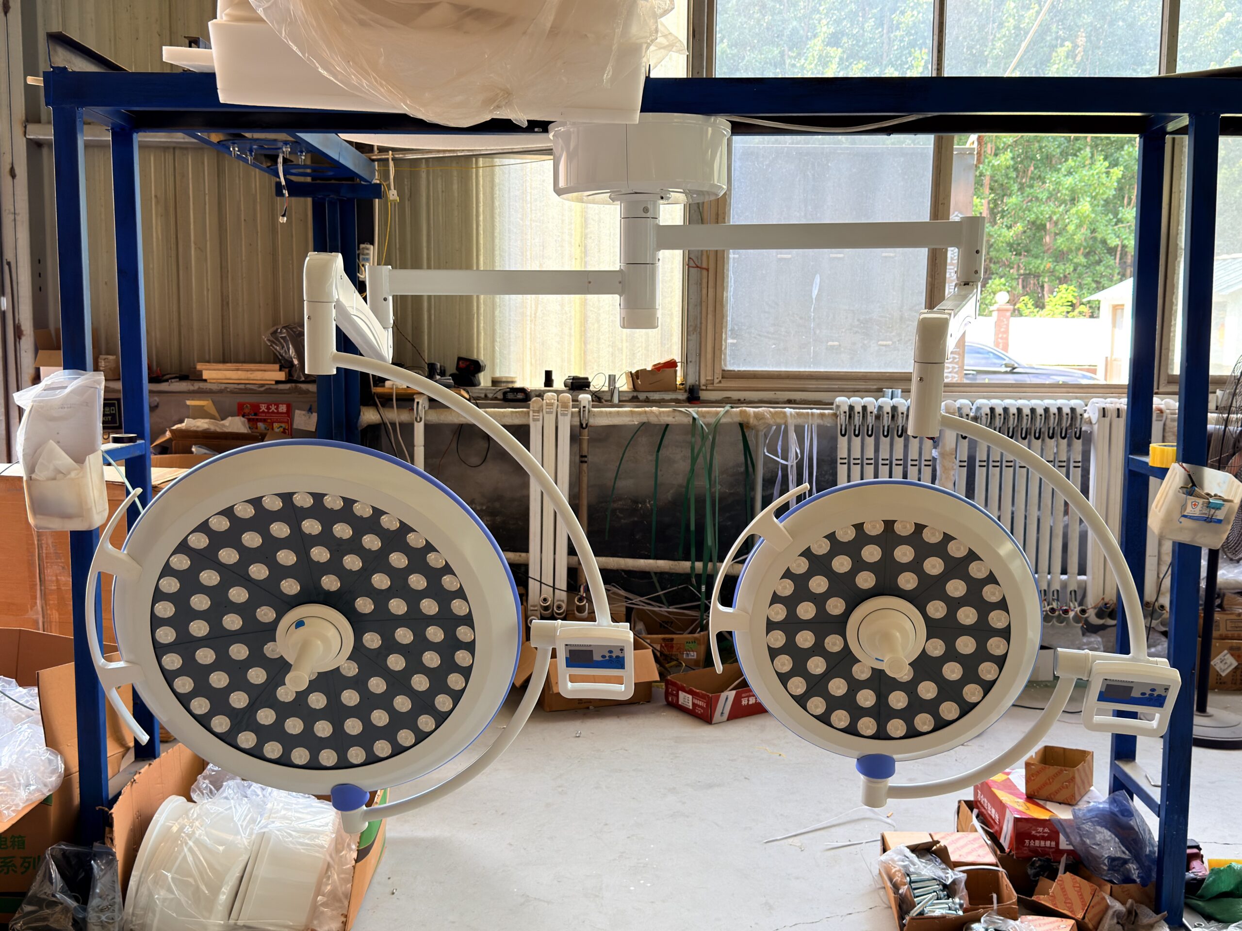
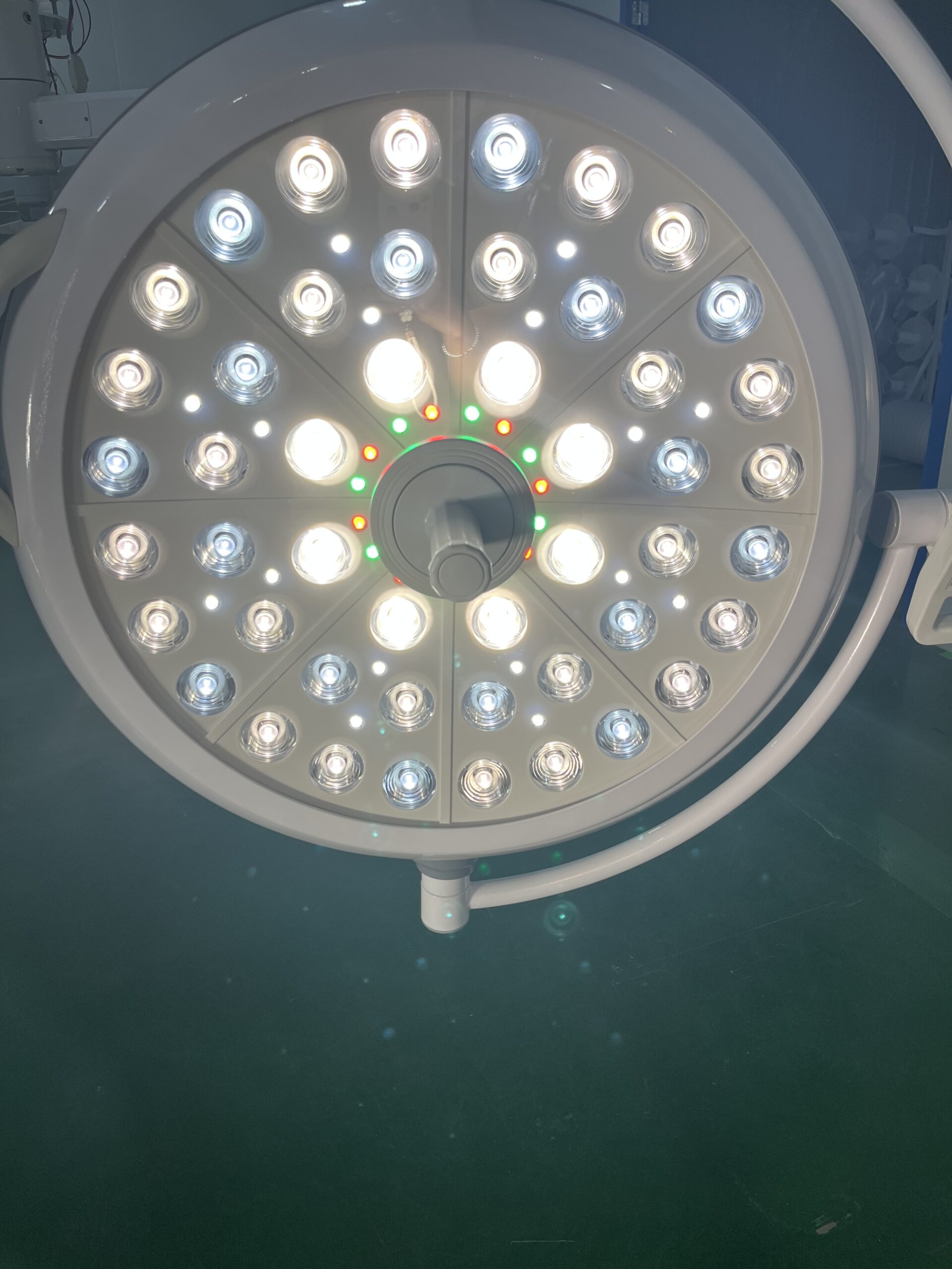
A bare LED is a small, intensely bright point — a near-ideal glare source. Left undiffused, an array of them produces a field full of hot spots and hard shadows. The first lever is to spread the same total output over a larger emitting surface, which lowers the surface luminance that drives direct glare. This is why large-diameter and multi-petal heads feel softer than compact ones at equal lux: the apparent source is bigger and less intense per square centimeter.
The second lever is individual optics per LED. Rather than letting the diodes spill raw light, quality heads give each LED its own lens or reflector so every point source projects a controlled beam onto the field. The beams overlap and blend into one continuous, uniform field instead of a constellation of bright points. This multi-source overlap is also the mechanism behind good shadow dilution: when the surgeon’s head blocks one set of beams, neighboring beams from different angles still reach the same spot, diluting the shadow rather than casting a hard one. Some manufacturers take this further with independently controlled modules and electronic field-size adjustment that changes the field diameter without moving parts.
- Larger, distributed emitting area lowers surface luminance for a given output, directly reducing direct glare.
- Per-LED lenses / reflectors turn point sources into controlled, overlapping beams, eliminating hot spots and producing soft field edges.
- Diffusing and micro-prismatic elements suppress the specular hot spots that cause veiling reflections off wet tissue and instruments.
- Multi-angle beam geometry gives strong shadow dilution — light arrives from enough directions that an obstruction never fully darkens the field.
- Independent module control (on advanced heads) tunes field diameter and balance electronically, keeping center intensity while softening the periphery.

Color Temperature, CRI, and Flicker: The Comfort Layer of Glare Control
Glare is mostly about luminance, but visual comfort is not. Three “softer” specifications interact with glare to determine how tired the team feels at hour four, and they belong in the same procurement conversation.
Color temperature. IEC 60601-2-41 constrains surgical lights to a correlated color temperature between 3,000 K and 6,700 K. A controlled study on LED lighting and eye strain (Ophthalmic and Physiological Optics, 2022) found that a moderate combination — around 500 lux at 4,000 K — produced the least asthenopia, while the most straining condition was high illuminance paired with a very cool 6,500 K. Separately, surgeons evaluating adjustable-color surgical light in a 2019 tissue study rated spectra around 4,000 to 5,100 K as highly effective, with 3,000 K rated less effective. A neutral, mid-range color is the comfort sweet spot, and adjustable color temperature lets surgeons dial in their preference.
Color rendering (CRI). High color rendering — a high Ra plus a strong R9 (saturated red) value — lets the surgeon distinguish oxygenated from deoxygenated tissue. It interacts with glare: when color rendering is poor, the team compensates by brightening the field to recover contrast, which then drives glare. Specifying strong CRI is therefore an indirect glare-control measure. Our article on surgical light color rendering degradation covers how CRI drifts over the life of the LEDs.
Flicker. Cheap LED drivers pulse the light at mains or switching frequency. The eye may not see it, but flicker contributes to headaches and fatigue over long shifts and can create stroboscopic artifacts on moving instruments and camera feeds. Modern surgical and ambient OR lighting targets flicker-free performance, with metrics such as PstLM at or below 1 and SVM at or below 0.4 increasingly cited in healthcare guidance aligned with EN 12464-1.

Visual comfort is a system property, not a single spec. A head with perfect optics but a harsh 6,500 K color, weak red rendering, and a flickering driver will still tire the team. Specify luminance control, color, CRI, and flicker together — or accept that you are solving a quarter of the problem.
Specifying and Verifying Glare Control at Procurement
Everything above is useless if it stays theoretical. Glare disappointments survive into commissioned operating rooms because comfort rarely becomes a verifiable acceptance criterion. The fix is to write glare control into the tender as testable line items and verify them on the installed head, not just on the brochure.
- Demand the full IEC 60601-2-41 ed.3 photometric set, not just peak lux: central illuminance at DRef, d10 and d50, depth of illumination L1 + L2, and shadow dilution — all at steady state after one hour at full intensity.
- Ask for the luminance distribution of the emitting surface — a uniform, low-peak-luminance surface is the physical signature of a low-glare head.
- Require smooth, wide-range dimming so the team can back off surface glare without the field collapsing or shifting color.
- Specify color temperature range and adjustability, plus CRI Ra and R9, and confirm a neutral, mid-range color with high red rendering.
- Request flicker data (PstLM, SVM) for the head and the ambient OR luminaires; treat flicker-free performance as a requirement.
- Get a UGR assessment for the ambient OR package at your actual room geometry and reflectances, confirmed against the relevant EN 12464-1 limit.
- Verify on site at commissioning: lux meter at field center and at depth, a shadow-dilution check, and a surgeon looking toward the head and monitors from normal working positions.
| Procurement Check | Document to Request | On-Site Verification |
|---|---|---|
| Deep-cavity reach & homogeneity | IEC 60601-2-41 report: EC,Ref, d10/d50, L1 + L2 | Lux meter at center and at depth; confirm soft field edge |
| Shadow handling | Shadow dilution data (mask and 50 mm cavity) | Obstruct part of the head; confirm field stays usable |
| Direct glare from the head | Emitting-surface luminance map / photometric file | Surgeon looks up at head; no harsh hot spot |
| Reflected glare & monitor wash-out | Optics description; ambient UGR study | Check reflections off instruments and screens |
| Color comfort & dimming | CCT range, Duv, CRI Ra/R9, dimming, flicker | Dim through full range; no color shift or flicker |
One final habit saves a lot of grief: treat operating room lighting as one coordinated system — surgical heads, ambient luminaires, and displays — rather than three unrelated purchases. The glare that fatigues a team is frequently born at the boundary between them. For a full theatre build or renovation, our turnkey operating room solution team lays out the surgical lights, ambient lighting, pendants, and displays as one integrated plan, and our surgical light maintenance and TCO guide covers keeping that glare performance stable over the life of the installation.
Conclusion
Surgical light glare control is the difference between a light that measures well and a light the team can work under for an entire day. It is not about reducing brightness — modern heads have brightness to spare — but about distributing it intelligently: a large, evenly diffused emitting surface; per-LED optics that blend into one uniform field; strong depth of illumination and shadow dilution; and a neutral color, high red rendering, and flicker-free drive that take the strain off the eye over long cases.
The standards give you the vocabulary — UGR for the ambient installation, IEC 60601-2-41 for the surgical head — but neither replaces verifying the answers on the installed equipment. Build glare control into your tender as testable line items, and check for direct and reflected glare at commissioning with a surgeon actually standing at the table. If you would like to discuss glare-controlled surgical lighting for a specific room or a full theatre program, talk to our engineering team — we would rather help you specify it correctly than fix it after the ceiling is closed.
Frequently Asked Questions
What is a good UGR value for operating room lighting?
UGR runs on a practical scale from about 13 to 28, with roughly three points the smallest noticeable step. EN 12464-1 sets limits around UGR 19 for patient and examination tasks — a sensible target for ambient operating room lighting. UGR applies to the room’s general lighting, not directly to the surgical head, which is governed by IEC 60601-2-41.
Why does a brighter surgical light sometimes feel worse, not better?
Glare is driven by luminance contrast and reflections, not total light alone. Concentrating energy at the tissue surface creates hot spots and specular reflections, and a bright field in a dim room forces constant pupillary re-adaptation. Good depth of illumination and a soft field edge let the surgeon see into the cavity at a lower, more comfortable surface brightness.
How does LED array design reduce glare?
Quality heads spread output over a larger emitting surface to lower surface brightness, and give each LED its own lens or reflector so the beams overlap into one uniform field with soft edges. That multi-source geometry also produces strong shadow dilution, reducing the temptation to over-brighten.
Does color temperature affect surgeon eye strain?
Yes. Surgical lights are constrained to roughly 3,000–6,700 K by IEC 60601-2-41, and a neutral mid-range color around 4,000–5,000 K tends to be most comfortable, while very cool white at high illuminance is linked to more eye strain. High color rendering and flicker-free drive also reduce fatigue.
How do I verify glare control before accepting a surgical light?
Make it a testable acceptance criterion. Request the full IEC 60601-2-41 ed.3 photometric set, emitting-surface luminance data, dimming range, color and CRI figures, and flicker metrics. Then verify on the installed head at commissioning with a lux meter and by having a surgeon check for direct and reflected glare from normal working positions.
You May Also Like...

Surgical Light Control Systems Explained: Touch Panels, Wall Controls, Sterile Handles and Integrated OR Control
A practical guide to the five surgical light control systems - touch panels, wall control...
Surgical Light Tender Specifications: The Complete Procurement Checklist
A practical checklist of the photometric parameters, certifications, and evaluation steps that belong in every...

Surgical Light Installation Requirements: Ceiling Load, Height and Coordination Guide
A practical guide to surgical light installation requirements: ceiling load and moment, clear height, arm...